White-nose Syndrome (WNS) has been decimating eastern Canada’s bat populations for the past six years. White-nose fungus, which thrives at low temperatures, often leads to hibernating bats waking up, flying into the cold, and freezing to death. In Canada, WNS was first discovered in Ontario and Quebec in 2009. In the Maritimes, the disease first appeared in New Brunswick and in Nova Scotia in 2011, and Prince Edward Island in 2013. The situation has grown dire; while NBM Zoologist Dr. Donald McAlpine and NBM Research Associate Karen Vanderwolf once found approximately 7,000 bats a year in the 10 hibernation sites in New Brunswick that they monitor, they found only 20 bats in the same caves last year. WNS affects primarily the Little Brown Bat and the Northern Long-ear Bat, although Big Brown Bats are also affected to a lesser extent. The decline in the bat population is expected to carry financial repercussions for agriculture and forestry as fewer bats will be consuming fewer crop and tree-damaging pests.
NBM Zoology Summer Students Maddie Empey, Alyson Hasson, and Neil Hughes have been working this summer to prepare and catalogue some of the approximately 7,000 Little Brown , Northern Long-eared , and Big Brown Bats from Ontario, Quebec, and the Maritime provinces in in the NBM freezers. These bats were all submitted by members of the public for rabies testing to a federal lab in Ottawa between 1996 and the early 2000s, before WNS was discovered in Canada. The bats at the NBM are those that tested negative for rabies.
The data collected from these bats will enable researchers to compare genetic variation in eastern Canadian bats before and after the introduction of WNS to the region. Among surviving bats, for example, there may be certain similarities in genetic makeup. Other research may use samples of fur to determine the levels of toxicants, such as mercury, that have been acquired by bats from the environment.
“This is a unique sample, in that it is probably the largest collection of those bat species most heavily impacted by WNS taken immediately before onset of the fungal infection,” said McAlpine. “Once archived in the NBM these samples will be a source of research data for many, many, years.”
“It’s really satisfying to know that you’re contributing to such research,” said Maddie Empey.
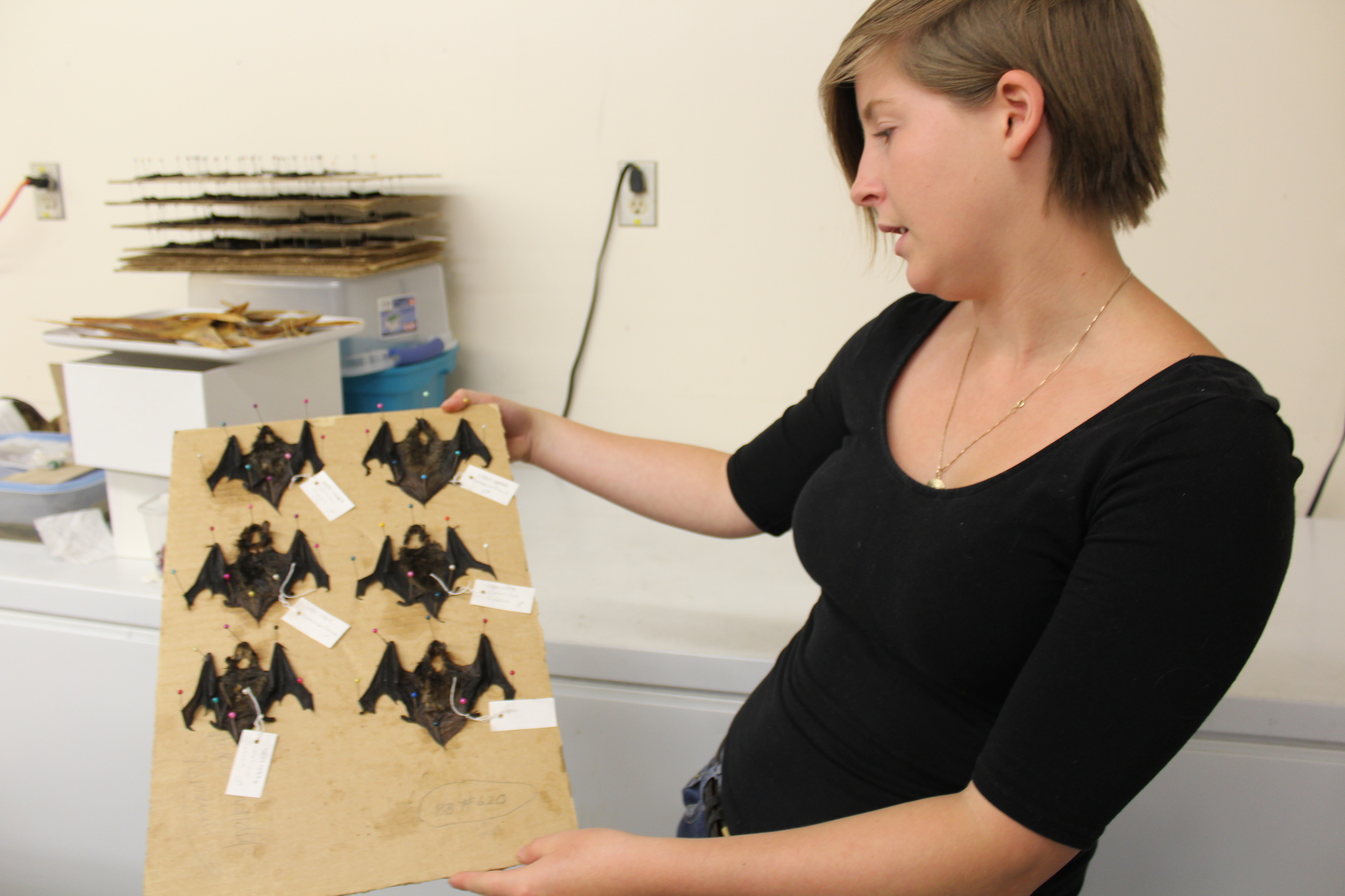
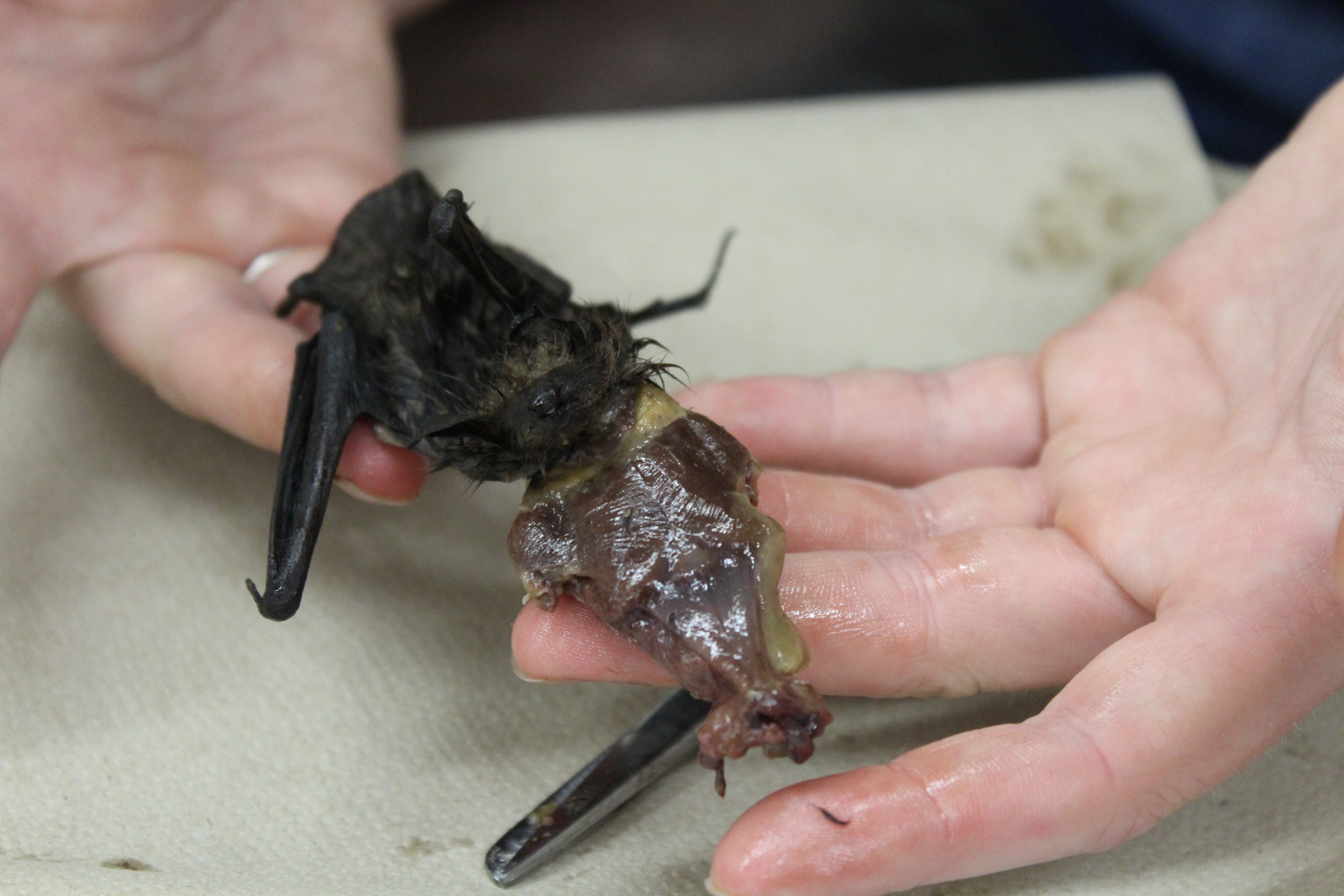
NBM Summer Student Maddie Empey holds samples of skinned bats.
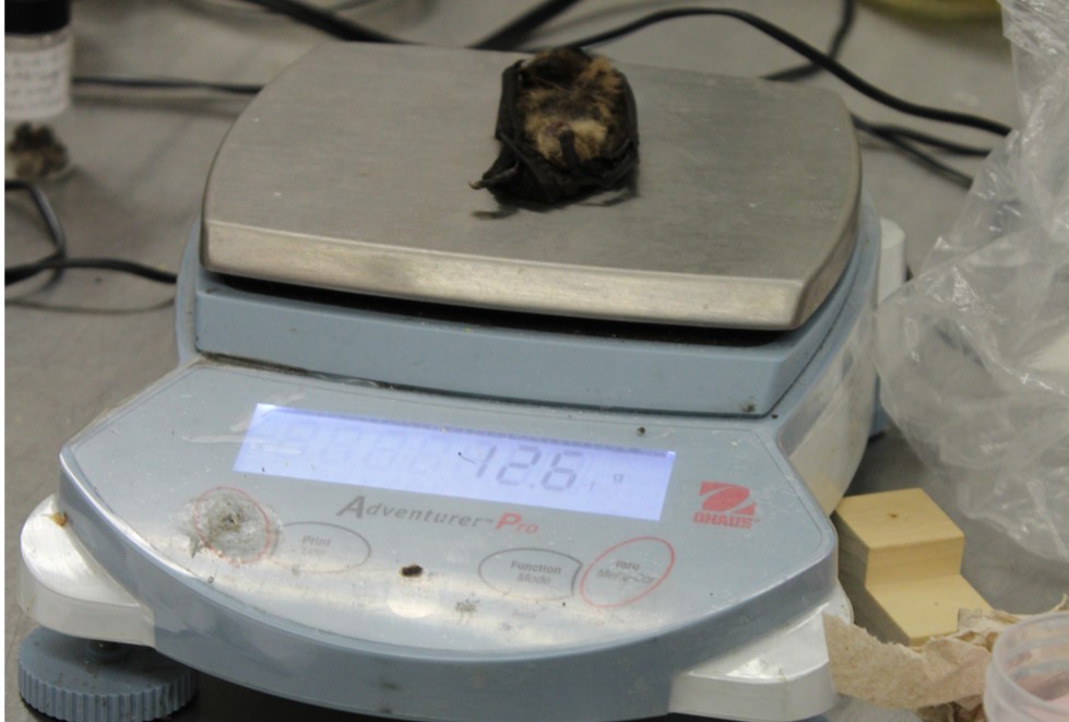
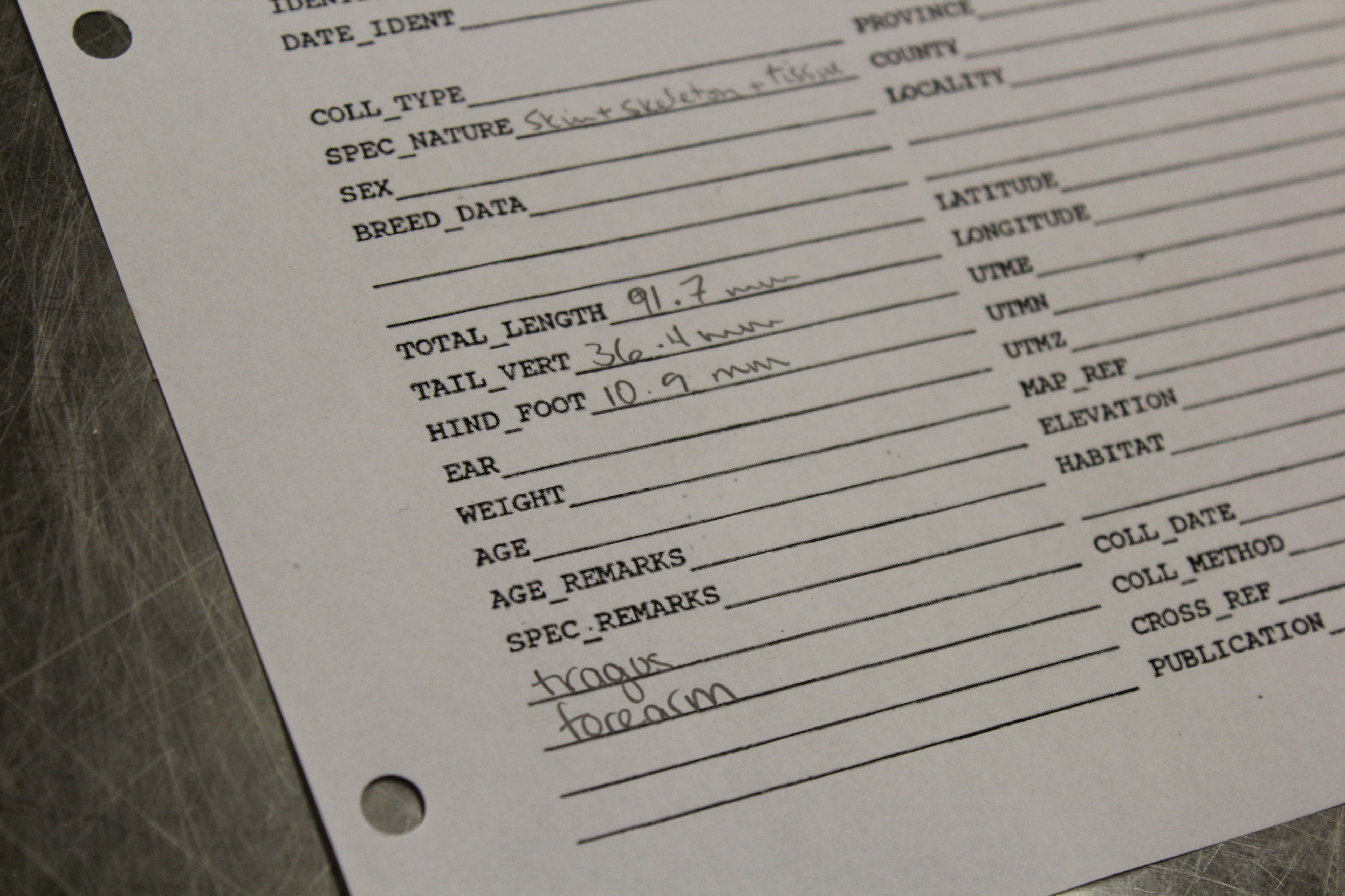
Students start by measuring the bats. Measurements include the length of the whole body, the tail, the hind foot, the forearm, and the tragus (a flap of skin in the ear involved in echolocation). Each bat is also weighed.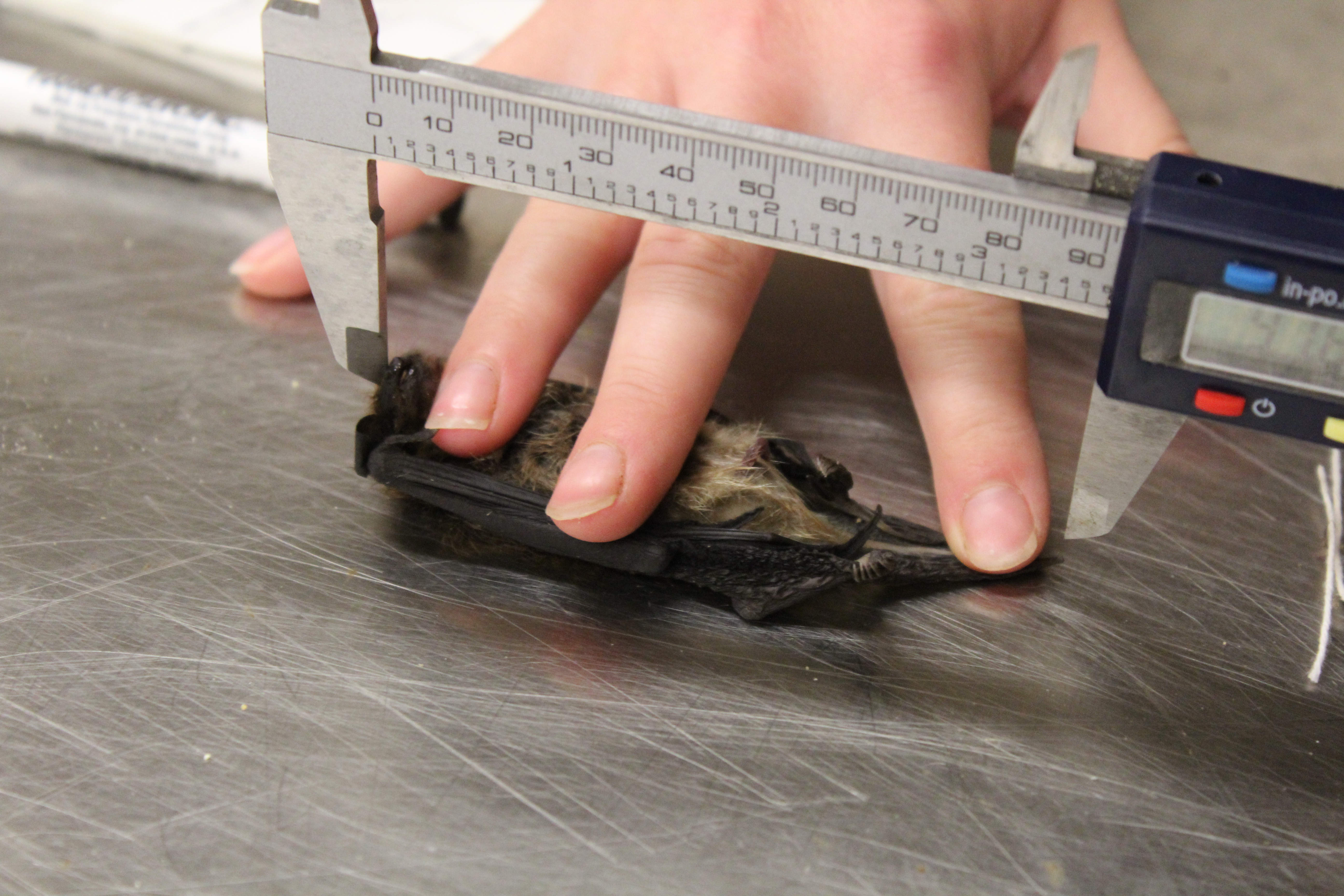
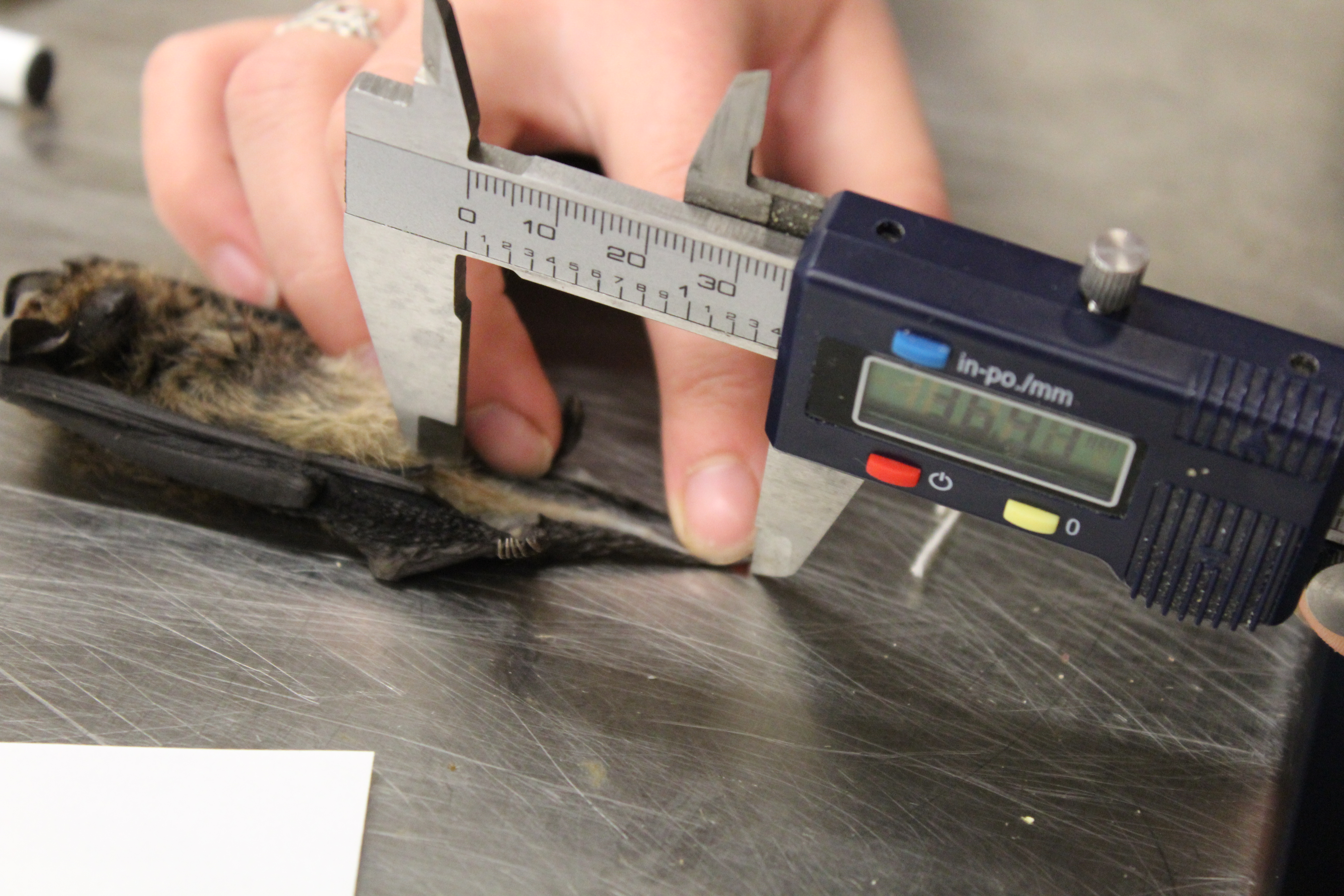
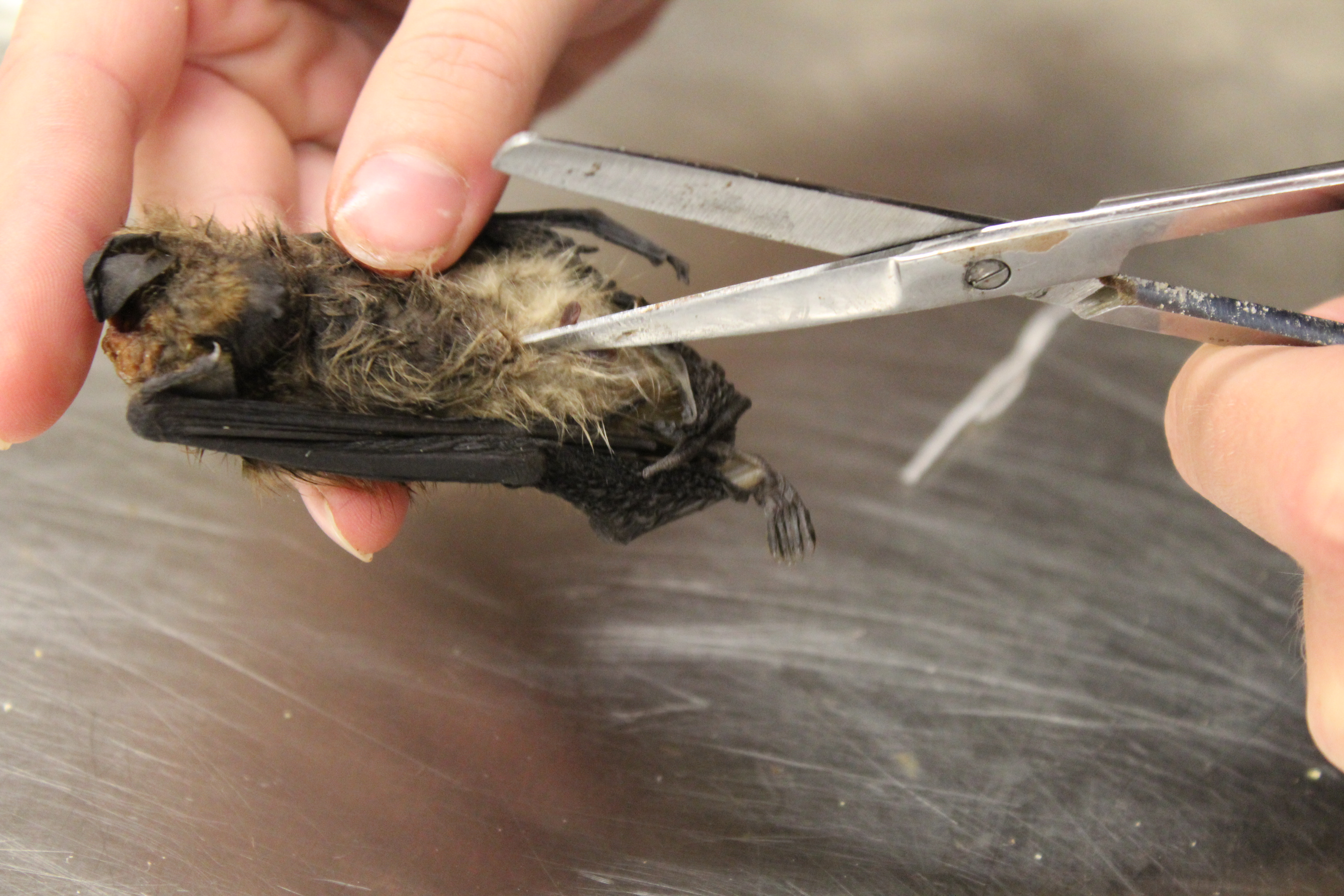
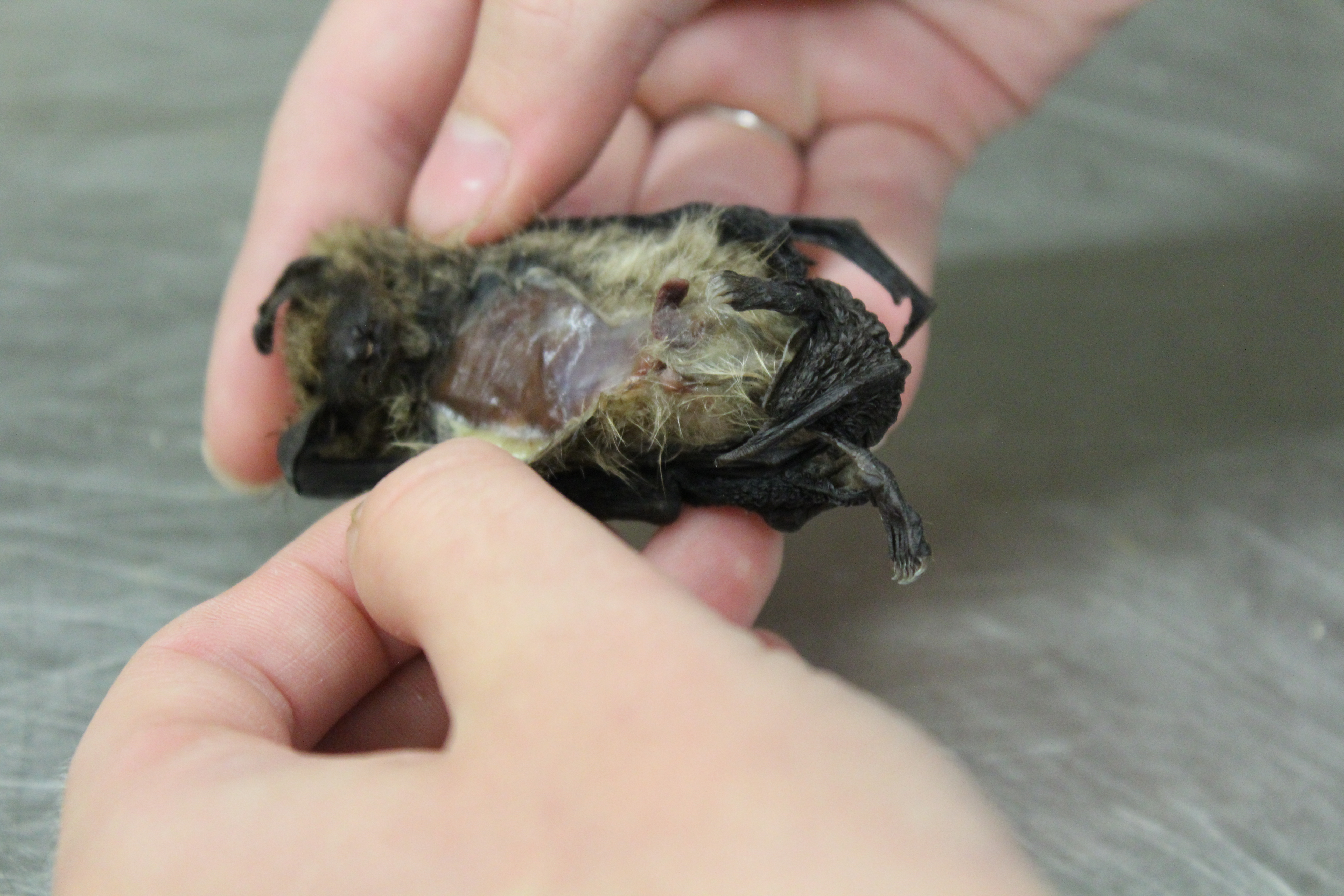
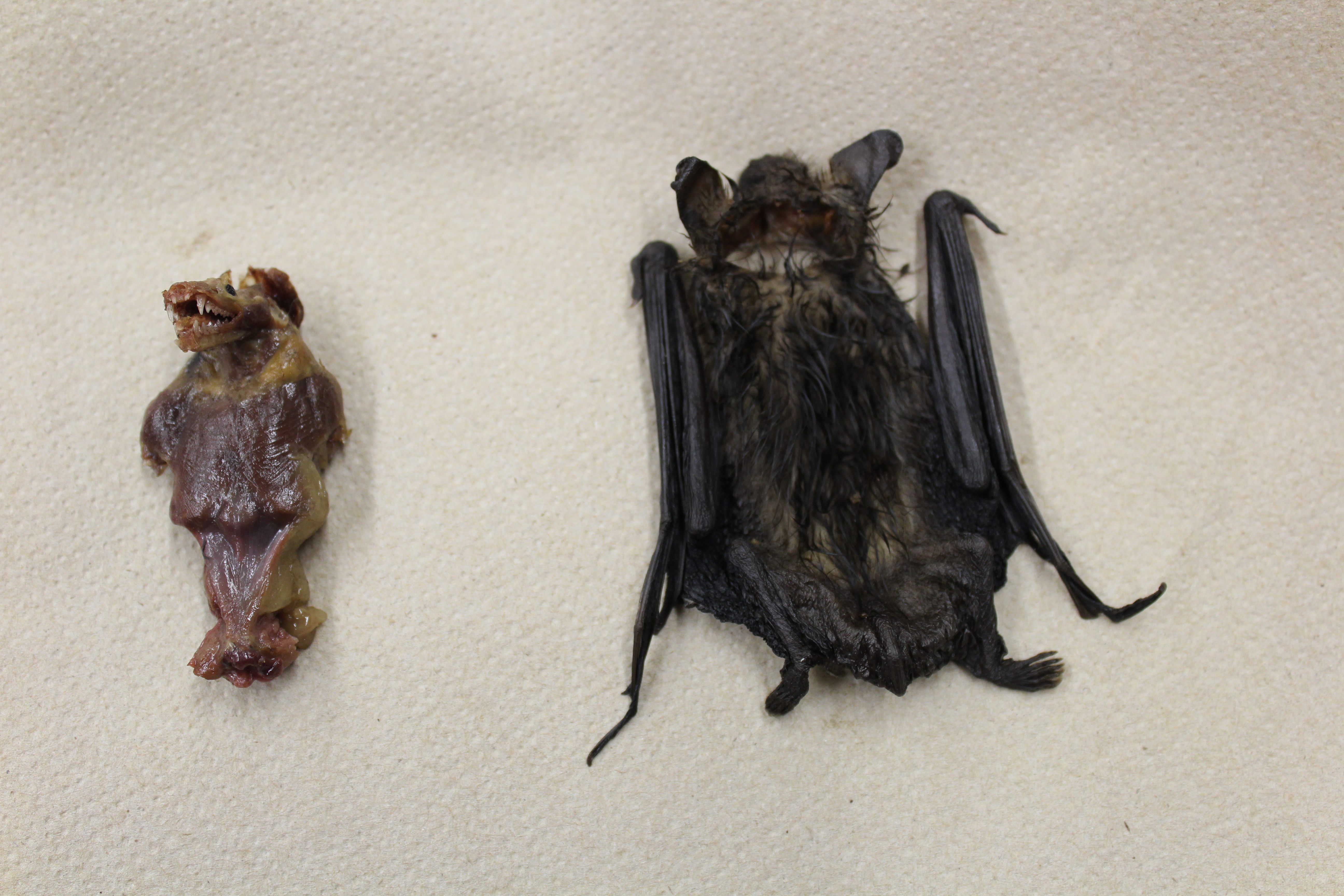
The bat skin is separated from the body. Although the wing bones remain with the skin, the remainder of the skeleton is retained for later cleaning and preparation.
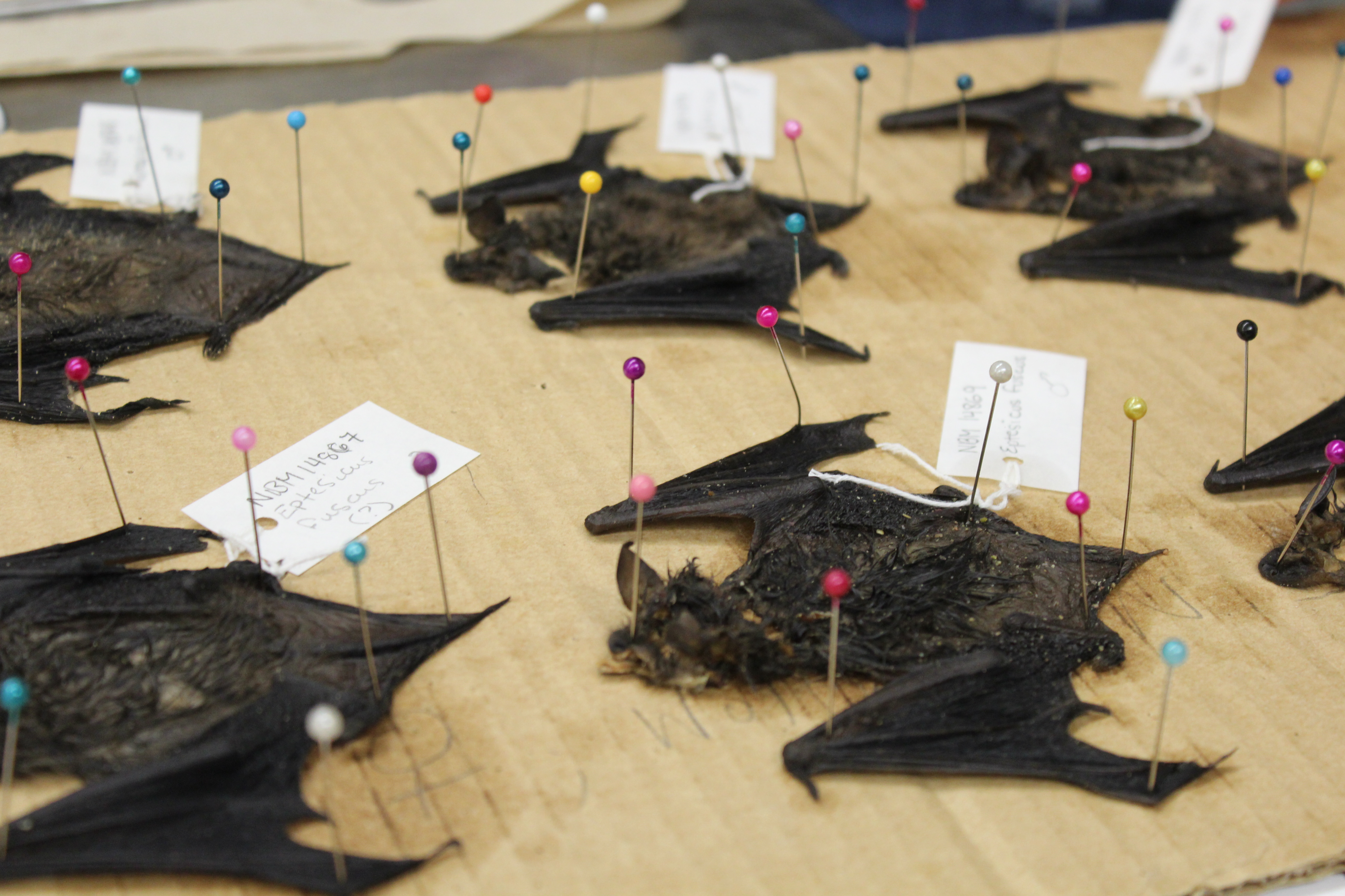
Each bat skin is spread and pinned to dry. Once dry, the skin will be placed in a clear Mylar envelope, and stored for future reference.
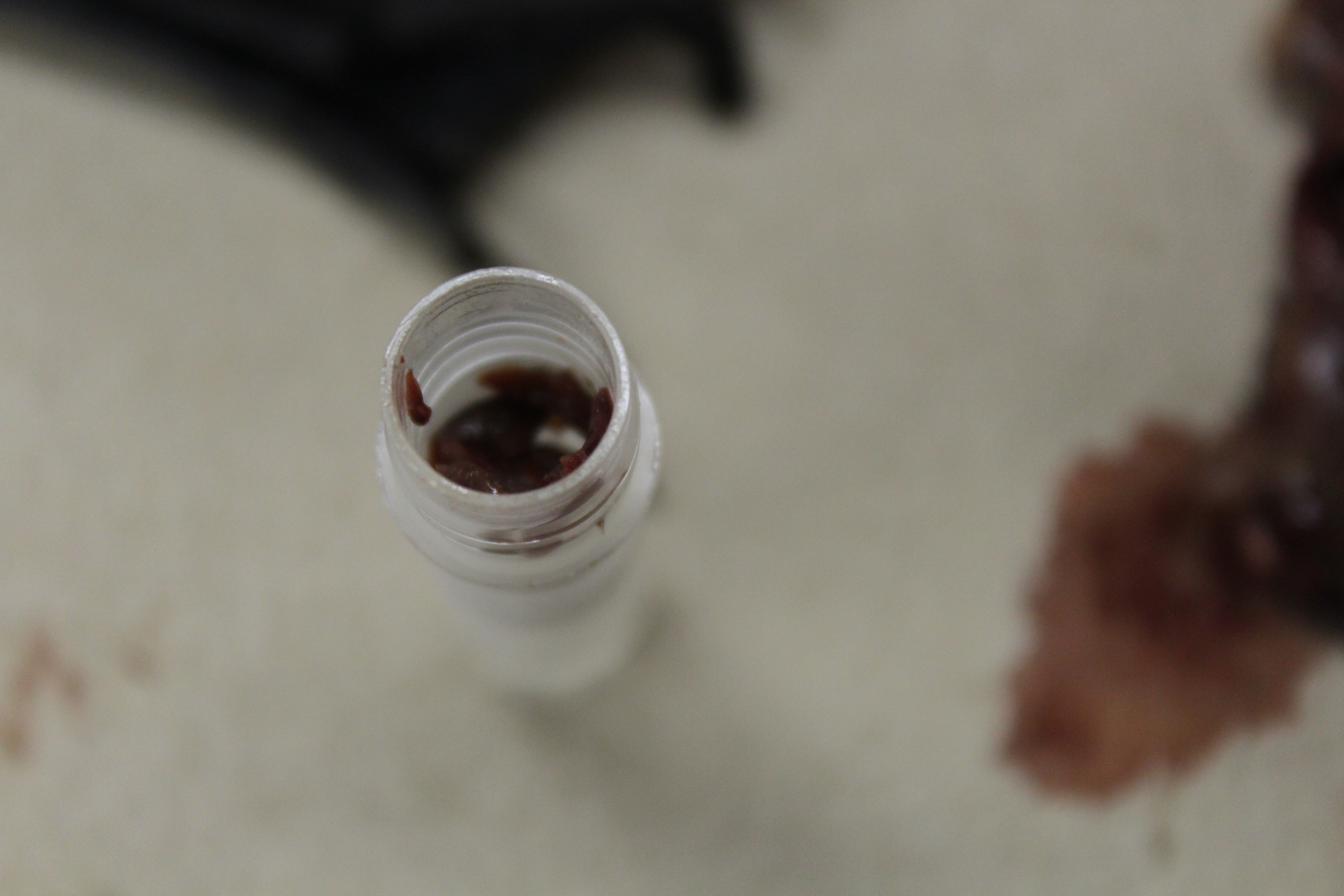
Tissue samples—small bits of muscle—are removed from each bat carcass, placed in 98% ethanol and stored in a freezer. Tissue samples from each bat are archived at -80o C in the NBM tissue collection for eventual genetic analysis by an NBM research collaborator at Trent University.
Finally, bat carcasses are placed in the dermestid beetle colony—or “bug barn” to be skeletonized. The beetles eat the flesh only, leaving perfectly cleaned skeletons. Once cleaned by bugs, the skeletons are removed, frozen, thawed, and frozen a second time to make sure that no beetles, eggs, or larva make their way into the NBM.
“If any beetles come in here [the NBM] they’ll just eat anything and everything,” said Empey.
Once the skeletons are cleaned and frozen, they are ready to be archived in the NBM collection to be used as reference for research.
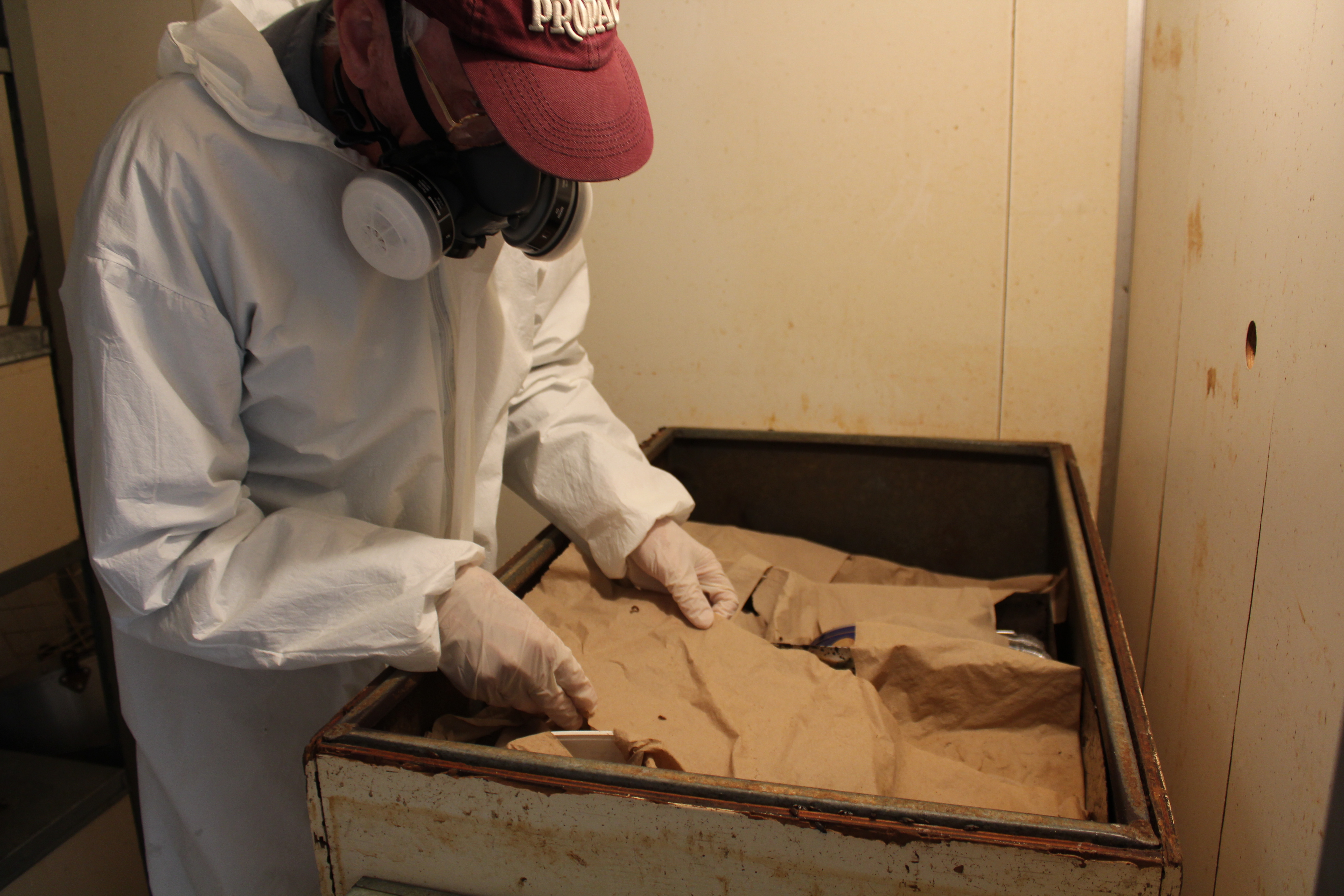
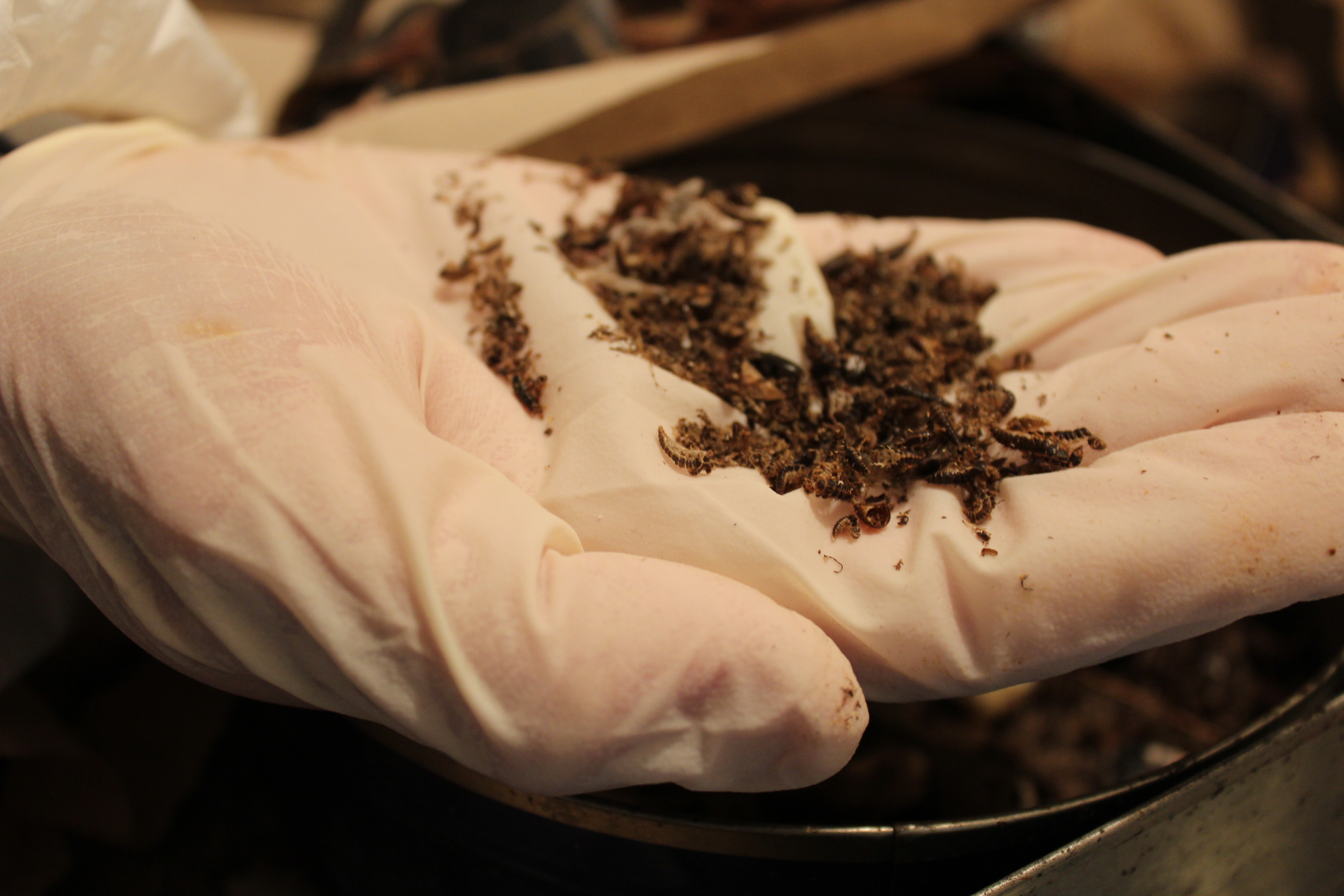
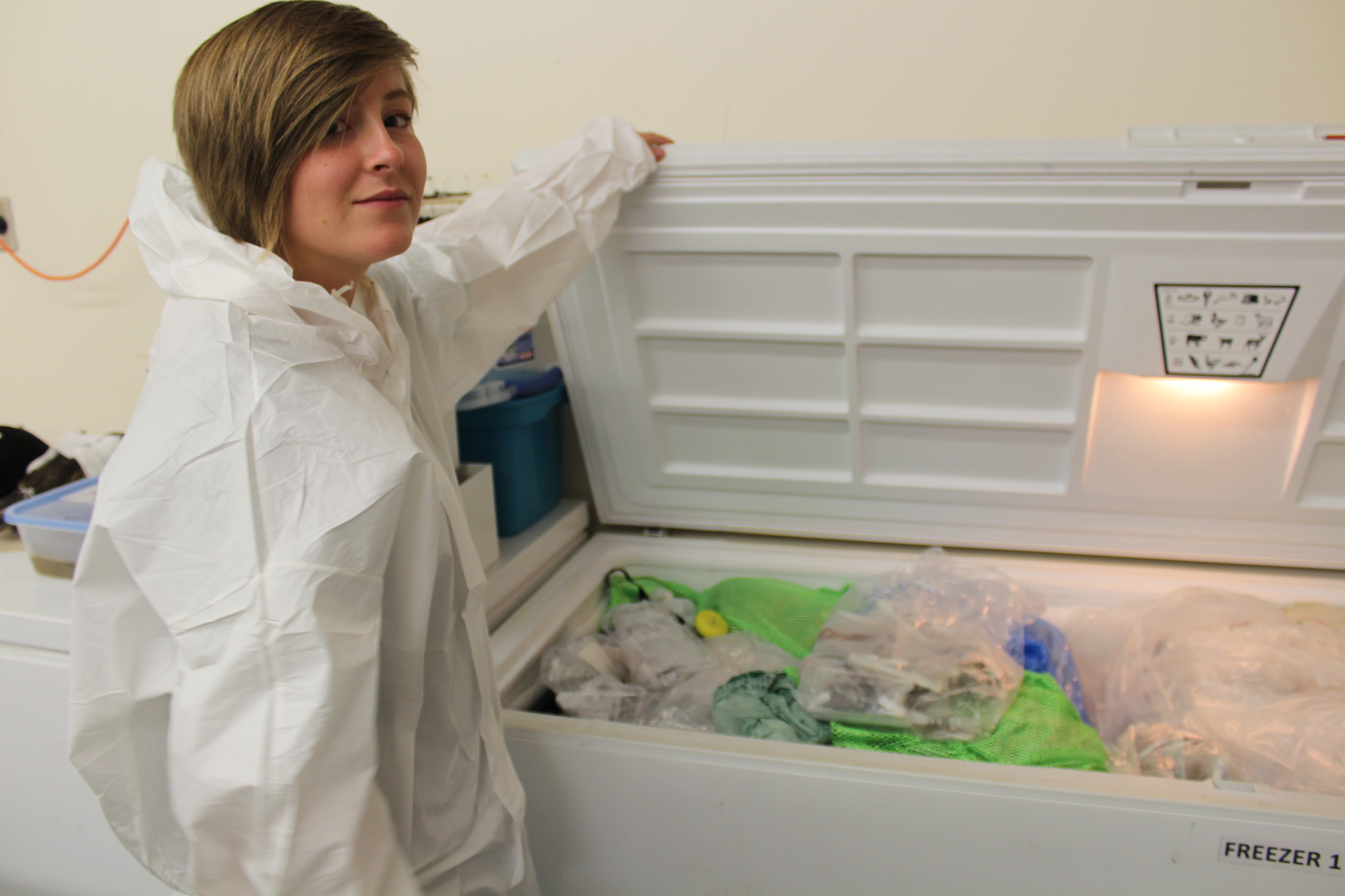
Clockwise from top left: NBM Preparator Brian Cougle with dermestid colony; Cougle holding dermestid beetle larvae; Empey with a freezer in the NBM necropsy lab.

